Eighteen years of published results.
Every paper published on IRIS technology, from the first in 2008 to the most recent.
journals, from PNAS to Optics Letters
41 venues in all, counting books and proceedings.
3,472 citations.
The ten most-cited papers in the library. Counts from Google Scholar, retrieved on October 12, 2026.
An additional 1,896 citations across the other 65 papers. the full library
Start here.
Eight papers, chosen to trace one line of work rather than to show the eight best results: the founding result, the sensitivity milestones, one component innovation, and a recent clinical application.

Label-free and dynamic detection of biomolecular interactions for high-throughput microarray applications

Digital detection of exosomes by interferometric imaging

Digital microarrays: Single-molecule readout with interferometric detection of plasmonic nanorod labels

Beating the reaction limits of biosensor sensitivity with dynamic tracking of single binding events
Attomolar sensitivity microRNA detection using real-time digital microarrays

Single virus fingerprinting by widefield interferometric defocus-enhanced mid-infrared photothermal microscopy

A spatially uniform illumination source for widefield multi-spectral optical microscopy

A label-free optical biosensor-based point-of-care test for the rapid detection of Monkeypox virus
The full library.
All 75, every one with a DOI that resolves. Nothing here sits behind an email capture.
2025
A label-free optical biosensor-based point-of-care test for the rapid detection of Monkeypox virus
Summary
Stopping an outbreak such as Mpox depends on tests that pair laboratory-grade sensitivity with the low cost and simplicity of a point-of-care device. The earlier single-particle form of the interferometric reflectance imaging sensor (IRIS) counted individual viruses on a silicon chip only after imaging each spot at a series of focus depths, a slow and costly step; the team's new Pixel-Diversity IRIS read each particle's optical signature from a single image instead. Capturing whole monkeypox virus on chip spots coated with Protein G and an anti-A29 monoclonal antibody (mAb 69-126-3), they detected it without labels down to 200 PFU/mL (plaque-forming units per milliliter, about 3.3 attomolar), against 1800 PFU/mL for a laboratory ELISA (enzyme-linked immunosorbent assay), and tests with herpes simplex virus type 1 and cowpox virus confirmed specificity. The authors see a path to point-of-care Mpox diagnosis, and the method's modular design could be adapted to test for several pathogens at once where high-affinity capture molecules exist.
2025
Impact of uniform illumination in widefield microscopy and mesoscopy
Summary
Widefield microscopes need even lighting to measure biological samples accurately at high throughput, and standard Köhler illumination often leaves large fields uneven. The team built a light source called EUCLID (effective uniform color-light integration device), and in two widefield imaging methods that depend on even light it gave significantly more precise quantitative readings than Köhler illumination. The authors expect it to suit many other widefield systems at low cost.
2024
Copolymer-Coated Gold Nanoparticles: Enhanced Stability and Customizable Functionalization for Biological Assays
Summary
Gold nanoparticles are prized in diagnostics for how they interact with light, but once loaded with antibodies, DNA or other biomolecules they can clump, stop working in complex biological samples, or stick to the wrong targets. The team coated citrate-stabilized and silica-shelled gold particles in copolymers based on N,N-dimethylacrylamide and checked them by light absorbance, nanoparticle tracking, electron microscopy and microarray tests. Two copolymers gave a stable coating with azide groups for attaching further molecules by click chemistry, and particles finished with single-stranded DNA and streptavidin worked in a biological assay. The method often needed just one step and a short incubation, and the authors note it can be tailored to different assays.
2024
Characterization of Receptor Binding Affinity for Vascular Endothelial Growth Factor with Interferometric Imaging Sensor
Summary
Wet age-related macular degeneration is the most common cause of sight loss in industrialized countries, and its biologic drugs work but are costly, so measuring how tightly each one grips its target helps confirm efficacy and compare options. The team printed two of them, the antibody bevacizumab and the fusion protein aflibercept, on one interferometric reflectance imaging sensor (IRIS) chip and watched in real time as recombinant human VEGF (vascular endothelial growth factor), which both drugs block, bound to them. Aflibercept bound VEGF more tightly than bevacizumab did, matching earlier results from two standard lab methods. The authors suggest IRIS, with low-cost silicon chips that run many measurements at once, is a promising tool for developing biologic drugs.
2024
Interferometric reflectance imaging sensor for diagnosis and therapy
Summary
For the 2024 Elsevier book Biophysics at the Nanoscale, Monireh Bakhshpour-Yucel, M. Selim Ünlü and four co-authors wrote a chapter on putting the interferometric reflectance imaging sensor (IRIS) to work in diagnosis and therapy. IRIS detects molecules captured on a silicon chip coated with a thin layer of silicon dioxide by the small change they make in the light the chip reflects, so the targets need no fluorescent or other label, and one chip can carry many capture spots read at the same time.
2023
Single virus fingerprinting by widefield interferometric defocus-enhanced mid-infrared photothermal microscopy
Summary
Standard virus tests cannot show what an individual virus particle is made of, since each holds so little: a vesicular stomatitis virus carries about 5000 proteins. The team built a widefield interferometric microscope that fingerprints many single viruses at once by how their chemical bonds absorb mid-infrared light. It identified protein and nucleic acid content in individual vaccinia and vesicular stomatitis viruses, told DNA from RNA viruses by thymine and uracil signals, and found extra β-sheet structure in varicella-zoster virus proteins. The authors suggest it could help analyze viral vectors.
2023
A spatially uniform illumination source for widefield multi-spectral optical microscopy
Summary
Uneven light skews widefield biological imaging, since real optics leave brightness and color patchy across the field. The team designed EUCLID (Effective Uniform Color-Light Integration Device), a simple, low-cost source that mixes light from separate emitters in an adjustable, diffusely reflecting hollow cavity, reshaping it to offset unevenness from the rest of the microscope. In multi-color imaging with either Nelsonian or Köhler alignment, it held brightness within about 1% across a wide field of view, a clear gain over the conventional method.
2023
Solid-phase optical sensing techniques for sensitive virus detection
Summary
Viral outbreaks can overwhelm health systems and disrupt daily life, and the standard clinical test, polymerase chain reaction (PCR), showed its limits during COVID-19 with slow turnaround and a need for sophisticated lab equipment. The authors surveyed optical biosensors that capture viruses on a solid surface, an approach valued for high sensitivity and a direct readout, covering fluorescence sensors, surface plasmon resonance, surface-enhanced Raman scattering, optical resonators and interferometric platforms. They then turned to their own group's single-particle interferometric reflectance imaging sensor (SP-IRIS), able to see individual nanoparticles, and showed how it detects viruses digitally by counting them one at a time.
2023
A high-throughput single-particle imaging platform for antibody characterization and a novel competition assay for therapeutic antibodies
Summary
Antibodies are central to diagnosing and treating Ebola, Sudan and Lassa virus infections, and choosing them takes large-scale screening. With the single-particle interferometric reflectance imaging sensor (SP-IRIS), which counts individual viruses caught on a chip, the team screened 30 monoclonal antibodies against stand-in viruses (recombinant vesicular stomatitis virus carrying each pathogen's surface glycoprotein) for the best capture antibodies, and used the binding patterns to map where each one attaches. Antibodies to the glycan cap or mucin-like domain of the Ebola glycoprotein gave the strongest signals, antibodies earlier shown to be highly effective in non-human primates; for Lassa, an antibody called 8.9F worked best and targets a region unlike the other 15 it was compared with. A new competition assay suggested why the ZMapp cocktail is more effective than two other Ebola cocktails: its three antibodies (13C6, 2G4, 4G7) did not block one another, and 13C6 even boosted binding by the other two.
2023
Bond-selective full-field optical coherence tomography
Summary
Optical coherence tomography (OCT), a label-free 3D imaging method widely used in research and clinical diagnosis, shows a sample's structure but not its chemistry. The team added a pulsed mid-infrared laser that heats specific chemical bonds, which modulates the OCT signal and yields 3D maps of where those bonds sit, even in samples that scatter light heavily. They showed it on 1 µm acrylic beads in agarose gel, polypropylene fibers from a surgical mask, cancer cell clusters, C. elegans worms and mouse brain tissue.
2023
Highly-sensitive, label-free detection of microorganisms and viruses via interferometric reflectance imaging sensor
Summary
Disease-causing microbes and viruses pass easily between hosts, and finding them fast, cheaply and without labels is key to prompt, appropriate treatment. The authors reviewed how the interferometric reflectance imaging sensor (IRIS) has been used to detect microorganisms and viruses in its two modes: at low magnification it measures the combined mass of molecules captured on each spot of the chip, and at high magnification it sees and counts single nanoparticles and viruses.
2022
Attomolar sensitivity microRNA detection using real-time digital microarrays
Summary
MicroRNAs, short RNA molecules whose levels have been linked to infections, heart disease and cancers, are attractive diagnostic markers, but standard DNA microarrays lack the sensitivity and specificity to measure them for diagnosis. The team built a multiplexed digital microarray that tags each captured microRNA with a gold nanorod and tracks targets arriving on the sensor in real time rather than reading once at the end, so rare targets stand out from plentiful background molecules that bind only weakly. With two microRNAs, miR-451a and miR-223-3p, they measured down to 100 aM (attomolar) and calculated a detection limit near 10 attomolar for each (under 1300 copies in a 0.2 ml sample), down from the femtomolar range of end-point reading, while total incubation shrank to 35 min from 5 h.
2022
Sensitive and real-time detection of IgG using interferometric reflecting imaging sensor system
Summary
Older techniques struggle to detect biomolecules quickly, cheaply and without labels. Using an interferometric reflectance imaging sensor (IRIS), the team measured the antibody immunoglobulin G (IgG) in water-based solutions, tracking binding in real time. Signal tracked concentration closely from 1 to 50 μg/mL (correlation coefficient above 0.94), with a detection limit of 0.25 μg/mL (1.67 nM), and the authors suggest the method could support direct biomarker detection in the clinic.
2022
The Effects of Three-Dimensional Ligand Immobilization on Kinetic Measurements in Biosensors
Summary
Drug developers need reliable measurements of how small molecules bind their targets, yet small molecules give weak signals on label-free biosensors, so sensor makers lean on surface chemistry, the way capture molecules are attached to the sensor, to make up the difference. Surface plasmon resonance sensors pack capture molecules into a thick dextran gel to boost the signal, but the analyte then has to diffuse through that layer, which can skew the measured binding strength; flat single layers built on epoxysilane avoid that, yet they demand extreme sensitivity and have been shown to make proteins unfold. The authors reviewed how these three-dimensional and flat attachment strategies affect binding measurements: stacked layers raise sensitivity but bring crowding, mass transport limits and other diffusion effects, while epoxysilane layers escape diffusion but can distort the capture molecules. They closed by weighing a hybrid that pairs a three-dimensional polymer coating with a single layer of capture molecules, setting out its strengths and weaknesses against the other two approaches.
2021
Vibrational spectroscopic detection of a single virus by mid-infrared photothermal microscopy
Summary
Chemical bonds warm slightly when they absorb infrared light, but in a single virus that signal is extremely faint. The team built a confocal mid-infrared photothermal microscope that uses interferometric scattering to detect it, and recorded label-free spectra from individual vesicular stomatitis viruses and poxviruses. Spectra were consistent within each virus type and dominated by two protein bands, amide I and amide II, whose ratio differed significantly between the two viruses, suggesting the method could tell viruses apart and, the authors propose, might help predict and control outbreaks.
2021
Background-Suppressed High-Throughput Mid-Infrared Photothermal Microscopy via Pupil Engineering
Summary
Wide-field mid-infrared photothermal microscopy identifies materials by the chemical bonds they absorb, but for particles smaller than the wavelength of light the faint signal is lost in the glare of reflected background light. The team reshaped the pupil of the microscope's collection optics to hold back most of that reflected light, cutting the background more than a thousandfold without losing sharpness and improving signal against background noise sixfold. They chemically told apart hundreds of nanoparticles in a 70 μm × 70 μm field, checked their imaging theory on 300 and 500 nm plastic beads, and imaged two bacterial species as well as fat droplets inside yeast and cancer cells.
2021
Configurable Digital Virus Counter on Robust Universal DNA Chips
Summary
Single-particle IRIS (SP-IRIS) counts individual virus particles as they land on a sensor chip, and the team built that chip on DNA rather than antibodies so it could be reconfigured. Antibodies tagged with DNA strands paired up with matching strands printed on the chip, forming an array that detected three model viruses carrying the surface proteins of Ebola, Marburg and Lassa, live in a disposable cartridge. A one-step version, with the tagged antibodies mixed into the sample first, detected the Ebola model at 100 PFU/ml within an hour, and in a pump-free cartridge at 104 PFU/ml in under 10 minutes, which the authors see as promising for point-of-care testing.
2021
The Role of Surface Chemistry in the Efficacy of Protein and DNA Microarrays for Label-Free Detection: An Overview
Summary
A microarray test is only as good as the capture molecules fixed to its spots: how densely they pack and whether they still work once attached. The authors surveyed common and recent ways of preparing chip surfaces to hold proteins or DNA, comparing how well each anchors them. They focused on label-free arrays that image single particles or measure binding kinetics, and weighed how polymer coatings affect the molecules they carry.
2021
Bond-selective interferometric scattering microscopy
Summary
Interferometric scattering microscopy images particles as small as proteins and viruses without labels but reveals only size and shape, while infrared spectroscopy reads chemical bonds yet cannot resolve individual nanoparticles. The team combined them: a pulsed mid-infrared laser heats particles that absorb at a chosen bond, and a visible beam reflecting off a layered thin-film chip detects the tiny change this makes in each particle's scattered light, read out by a camera acting as a lock-in detector. After tests on polymer beads smaller than the wavelength, they chemically imaged tens of particles at once, including E. coli, S. aureus and the yeast Candida albicans.
2021
A Reliable, Label Free Quality Control Method for the Production of DNA Microarrays with Clinical Applications
Summary
For DNA microarrays to be trusted in clinical routine, every spot must carry its capture probes, since a badly printed spot can weaken a test or cause a false negative. The authors used IRIS, which measures, without labels, how much material sits on the chip, to inspect chips right after printing. The check caught spots missing their probes, which later gave no signal in a clinically relevant fluorescence test, and found the probe concentration that maximized target capture before overcrowding set in. Comparing two attachment chemistries on two coatings in a colorectal cancer assay, they found that packing in more probe did not raise the binding signal, probably because of crowding.
2021
Multiplexed Affinity Measurements of Extracellular Vesicles Binding Kinetics
Summary
Extracellular vesicles, tiny membrane packets released by cells, carry clues about disease, but profiling them usually takes slow optimization and incubations that can run overnight. With IRIS in a flow chamber, the team watched vesicles bind a multiplexed chip in real time without labels, and in a single experiment of under an hour measured binding to 18 probe conditions: three antibodies, each printed at six concentrations. They fitted the data with a model mixing fast and slow rates of binding and release, since a large vesicle grips many probes at once, and confirmed that vesicles bound more strongly to the anti-CD81 antibody than to anti-CD9 or anti-CD63; single-vesicle imaging with fluorescent staining backed this up.
2021
Bulk-Effect-Free Method for Binding Kinetic Measurements Enabling Small-Molecule Affinity Characterization
Summary
Label-free optical sensors register binding as a change in refractive index, so a change in the liquid itself, from a solvent, temperature or pH, can masquerade as binding; surface plasmon resonance and biolayer interferometry leave this bulk effect uncorrected or subtract it with a reference channel. The team instead tuned the blend of LED colors lighting the IRIS sensor so that it barely responds to the bulk liquid, with no reference channel or software correction. Switching from buffer with a refractive index of 1.335 to buffer containing 1% of the solvent dimethyl sulfoxide (DMSO), at 1.336, moved the signal by only about 8 × 10⁻⁶, and the team cleanly recorded biotin, a 244.3 Da molecule at 1 μM in 1% DMSO, binding to streptavidin.
2020
Highly sensitive and label-free digital detection of whole cell E. coli with Interferometric Reflectance Imaging
Summary
Spotting disease-causing bacteria fast helps stop infections spreading, and sensors that catch whole bacteria directly skip the time and cost of labeling them or extracting their components. The team used interferometric reflectance imaging to see and count individual E. coli cells captured across a large sensor, and from buffer measurements with no sample preparation extrapolated a detection limit of 2.2 CFU/ml (colony-forming units, a count of live bacteria), which the authors believe is the most sensitive label-free detection of whole E. coli reported. The sensor picked out E. coli over three other bacterial species, worked as well in tap water, and its magnification let each detection be confirmed as the target by its shape. The authors see this as a route to testing real-world and clinical samples directly.
2020
High-throughput, high-resolution interferometric light microscopy of biological nanoparticles
Summary
Viruses and other biological nanoparticles are hard to see under a light microscope: they are tiny, barely stand out from the surrounding liquid, and blur below the resolution limit. Interferometric microscopy already solves the contrast problem, so the team added computational imaging, lighting samples asymmetrically and reconstructing particles with a rigorous optical model. This doubled lateral resolution, to about 150 nm, across a field larger than 100 × 100 μm2, imaging more than 104 particles at once without labels, and revealed the shapes of many Ebola virus-like particles as well as an Ebola vaccine candidate with a distinct structure.
2020
Highly multiplexed label-free imaging sensor for accurate quantification of small-molecule binding kinetics
Summary
Small molecules such as drugs and toxins add so little mass when they bind a protein that they are hard to follow without labels. The team lowered the detection limit of IRIS to track such binding in real time on an antibody microarray, reaching 1 pg/mm2 sensitivity in a biotin and streptavidin test by averaging over space and time. Screening 20 antibodies at once against fumonisin B1 (721.8 Da), a toxin common in corn and wheat, gave binding curves with signal about 8 times the noise.
2020
Computational nanosensing from defocus in single particle interferometric reflectance microscopy
Summary
In single-particle interferometric reflectance microscopy, each nanoparticle changes appearance in a distinctive way as the focus moves through it. The team turned that stack of defocused images into one clear, low-noise image per particle, using a fast, memory-light reconstruction built on a physical optics model, and tested it on silica beads of nominally 50 nm.
2020
Simultaneous evaluation of multiple microarray surface chemistries through real-time interferometric imaging
Summary
How well a chip's coating holds its capture molecules caps how much binding a microarray test can detect, so choosing the coating matters. The team printed many different reactive polymers as separate spots on one bare silicon/silicon dioxide chip, then deposited the same capture molecule onto every spot, so that dozens of coatings could be judged side by side in a single 60-minute experiment; the approach worked for proteins as well as short DNA strands. Real-time binding measurements with IRIS showed the printed spots were about as uniform as a conventional coating spread over the whole chip, and comparing binding curves across spots revealed which polymers best anchored a model protein, α-lactalbumin, and a peptide taken from it, LAC-1. Because each spot carried its own chemistry, the method let molecules with different chemical handles share one chip, each with its attachment tuned separately.
2020
Instrument-free protein microarray fabrication for accurate affinity measurements
Summary
Protein microarrays let researchers measure many binding interactions at once, but printing them usually takes expensive robotic spotters and trained operators, out of reach for many small labs. The team developed a low-cost way to make the arrays by hand with an ordinary micropipette, then compared hand-made and robot-printed spots of the protein α-lactalbumin by tracking how antibodies against it bound, using the IRIS platform. Hand-pipetted spots gave about 4-fold more binding signal, and a signal-to-noise ratio about 3 times better, than the average of 75 robotic spots covering the same sensor area. On a 24-test chip, they measured antigen-antibody binding constants with under 5% measurement error.
2019
Beating the reaction limits of biosensor sensitivity with dynamic tracking of single binding events
Summary
Ultrasensitive tests that take one reading at the end are limited by capture molecules that also cling, briefly, to plentiful background molecules. The team used gold nanorods as labels and interferometric reflectance imaging to watch and time thousands of individual binding events as they happened; true targets stayed bound longer. Across 12 tests on one sensor, they detected a synthetic DNA target at 19 fM.
2019
Interferometric Reflectance Imaging Sensor (IRIS) for Molecular Kinetics with a Low-Cost, Disposable Fluidic Cartridge
Summary
Picking binding partners for a diagnostic or therapy means knowing how they bind, which is hard to measure cheaply across many unmodified candidates. This methods chapter set out how to screen hundreds of unlabeled ligands at once with IRIS, using an inexpensive disposable cartridge that sits on the sensor and simplifies handling reagents.
2018
Digital microarrays: Single-molecule readout with interferometric detection of plasmonic nanorod labels
Summary
Microarrays read tens of thousands of DNA or protein targets at once, but their fluorescent readout is only modestly sensitive and spans a narrow concentration range, and earlier single-molecule versions needed amplifying chemistry or gave up speed. The team tagged each captured molecule with a gold nanorod and built an interferometric scanner that counts nanorods one at a time through a 10× objective, with no chemical signal boosting, at roughly the pace of a commercial fluorescence scanner. By counting particles in sparse spots and measuring dense spots as a whole, the system covered about 6 orders of magnitude of concentration in a single scan, with sensitivity and range each about 3 orders of magnitude beyond conventional arrays. In buffer, a proof-of-concept protein assay picked up hepatitis B virus surface antigen down to 3.2 pg/mL.
2018
Interferometric detection and enumeration of viral particles using Si-based microfluidics
Summary
Single-particle IRIS counts individual viruses without labels, which the group had already shown in cartridges built from laminated layers. They proposed a cheaper, compact cartridge made with widely available silicon manufacturing methods, designed to watch single viruses bind across a 9-spot array at once, simply and robustly enough for routine real-time use.
2018
Resolution enhancement of wide-field interferometric microscopy by coupled deep autoencoders
Summary
Viruses and exosomes lie below the resolution limit of light microscopes, so sharper images would help an interferometric microscope detect and classify them. The researchers trained a deep-learning network of coupled autoencoders on microscope images of L-shaped nanostructures paired with hand-made reference images. It then returned cleaner, higher-resolution versions of images it had not seen.
2017
Quantitative interferometric reflectance imaging for the detection and measurement of biological nanoparticles
Summary
Once captured on a protein microarray, single viruses and exosomes can be seen without labels by interferometric reflectance imaging, but their size and shape had been judged against reference particles of known size, an inconsistent, qualitative method. The authors explained the physics of how the sensor sees a particle and built an optical model of light scattering from particles of any shape, including rod-shaped and filamentous viruses, so the method can measure as well as detect. They then used it to tune the optics for small, weakly scattering particles in water.
2017
Pupil function engineering for enhanced nanoparticle visibility in wide-field interferometric microscopy
Summary
Biological nanoparticles scatter little light, so they are hard to pick out in a wide-field interferometric microscope. The team filtered light at the microscope's pupil plane, an approach called pupil function engineering, and checked the gains against theory. Polystyrene nanospheres about 25 nm in radius became ten times more visible, with a signal-to-background ratio of nearly 8%. The authors suggest the method could handle faint particles from several hundred nanometers down to a few tens, nearly the whole biological size range.
2017
Enhanced light microscopy visualization of virus particles from Zika virus to filamentous ebolaviruses
Summary
Ordinary light microscopes show bacteria, fungi and parasites, but unlabeled viruses are too small to see, so they are usually studied by electron or atomic force microscopy, which need purified samples and expert hands and count only a few particles per view. Using single-particle IRIS, the team captured virions straight from complex samples onto a silicon chip and automatically counted and sized thousands of them. Changing the light's color widened what it could see: violet/UV light revealed single flavivirus particles of about 40 nm, such as Zika virus, green light told vaccinia virus (about 360 nm) apart from vesicular stomatitis virus, and infectious ebolavirus and its virus-like particles showed their filaments clearly. The authors see a simple benchtop tool with uses from rapid virus detection in body fluids to vaccine work with virus-like particles.
2017
Disposable cartridge platform for rapid detection of viral hemorrhagic fever viruses
Summary
Individual viruses have been beyond the reach of light microscopes, so counting them has meant electron microscopy and purified samples. Aiming at hemorrhagic fever viruses, the team built a sealed, disposable cartridge around a multiplex interferometric imaging method, with capillary flow drawing sample across the chip at about 3 μl per minute so viruses could be watched binding, label-free, in complex samples such as serum. Binding showed within a 20-minute run, with sensitivity comparable to ELISA and plaque assays and equal to or better than paper-based point-of-need tests.
2017
Surface chemistry and morphology in single particle optical imaging
Summary
Sensors that capture whole viruses and exosomes without labels could simplify sample preparation for markers of conditions from infections to cancer, and may give better measurements because they keep information about each particle's nanoscale structure. Imaging sensors of this kind catch particles on a flat surface coated with capture molecules, and that coating is what makes them specific. It can also hold them back: because the sensors respond to tiny local changes in how the surface bends light, a rough or uneven coating adds noise. The authors reviewed label-free imaging approaches, comparing interference reflectance imaging with dark-field and surface plasmon resonance imaging, explained why smooth, uniform capture coatings matter, and summarized methods developed to produce them.
2017
Nanoparticle classification in wide-field interferometric microscopy by supervised learning from model
Summary
Interference-enhanced imaging can do more than spot particles below the resolution limit: how a particle's image changes as the focus shifts hints at what it is. The team trained a classifier on images simulated from a physical model of the microscope, then in blind tests used it to sort gold and polystyrene nanospheres by material and size.
2017
Visualizing ebolavirus particles using single-particle interferometric reflectance imaging sensor (SP-IRIS)
Summary
Seeing the long, threadlike form of Ebola virus normally takes an electron microscope. This methods chapter set out a light-microscopy alternative: intact Ebola virus and its virus-like particles were caught on a silicon chip printed with virus-specific antibody spots, then imaged by interference reflectance imaging, which registered each captured particle and resolved the filaments. Capture was possible from samples as varied as tissue culture medium and blood, without labels, and automated counts could give an unknown sample's virus concentration plus a quantitative view of particle sizes and shapes.
2017
Label-Free and High-Throughput Detection of Biomolecular Interactions Using a Flatbed Scanner Biosensor
Summary
Fluorescent microarrays are sensitive, but uneven probe attachment and poor repeatability weaken their numbers. The team turned an off-the-shelf flatbed scanner costing about 100 USD into a label-free interferometric reflectance imaging sensor for reading arrays. It tracked DNA strands pairing and antibodies anchoring through DNA; its noise floor was ~30 pg/mm2, and a 2 mm × 2 mm field took 5 s to scan (50 s with 10 frames averaged). The authors propose it for low-resource settings and for calibrating and checking fluorescence arrays.
2017
DNA-Directed Antibody Immobilization for Robust Protein Microarrays: Application to Single Particle Detection
Summary
Protein microarrays test one small sample for many targets at once, but only if the capture proteins stay attached and keep working. The authors showed how to build sturdy arrays by anchoring antibodies through DNA, with a strand tethered to each antibody pairing with a matching strand on the chip. Focusing on arrays that catch nanoparticles such as intact viruses, they used single-particle IRIS experiments to demonstrate the method's advantages.
2016
Digital detection of exosomes by interferometric imaging
Summary
Cells release exosomes, membrane-wrapped vesicles 30–100 nm across linked to cell-to-cell signaling, cancer spread and early disease detection, but their size and weak optical contrast make them hard to examine one by one. Using single-particle IRIS, the team captured exosomes on an antibody microarray chip, counted individual vesicles larger than 50 nm and sorted them by surface markers, first with purified exosomes from cultured HEK 293 cells. In human cerebrospinal fluid, antibodies against tetraspanins, proteins common on exosome surfaces, then captured exosome-sized particles from a sample of only 20 μL.
2016
Real-Time Capture and Visualization of Individual Viruses in Complex Media
Summary
Watching single viruses has usually required fluorescent labels or an electron microscope, both slow and expensive. The team combined single-particle IRIS with microfluidics so that single 100 nm vesicular stomatitis viruses could be counted live, without labels, as they bound to an antibody microarray. In undiluted fetal bovine serum, they captured and imaged rVSV-ZEBOV, an engineered vesicular stomatitis virus used as an Ebola model, at 100 PFU/mL (plaque-forming units per milliliter) in under 30 min.
2016
Physical modeling of interference enhanced imaging and characterization of single nanoparticles
Summary
Interferometric imaging is more sensitive than imaging scattered light alone. The authors built a physical model of how such microscopes image and characterize single nanoparticles, mapped what drives the signal, and checked it against experiments.
2016
Robust visualization and discrimination of nanoparticles by interferometric imaging
Summary
Counting captured particles one by one can detect far smaller amounts of a target than a bulk measurement, but only if faint particles are seen reliably. After surveying optical methods for spotting single nanoparticles, the authors described image capture and software changes to the single-particle interferometric reflectance imaging sensor (SP-IRIS) that sharply cut both missed particles and false counts, a step they called critical for bringing SP-IRIS into medical diagnostics.
2016
Numerical techniques for high-throughput reflectance interference biosensing
Summary
Turning raw spectral readings from the interferometric reflectance imaging sensor (IRIS) into binding measurements had been too slow for large studies, even though the technique suits tracking many molecular interactions at once. The team replaced the slow step with a precomputed lookup table, cutting processing time 10,000-fold while keeping results accurate. The table also worked with oxide thicknesses and instrument setups that fitting-based analysis could not handle.
2016
New approaches for virus detection through multidisciplinary partnerships
Summary
Viral outbreaks often strike where clinical laboratories are scarce, and sensitive tests tend to be costly and tied to those labs, a gap that cheap, portable tests could fill. The authors recounted developing a virus test that needs no label on the particle itself, aiming for speed and simplicity without losing sensitivity. They credited a team spanning many disciplines, which weighed candidate technologies, spun out a company and developed a diagnostic product for infectious disease.
2015
Digital detection of biomarkers assisted by nanoparticles: application to diagnostics
Summary
Tallying single molecules, rather than averaging a signal over many, is opening a new frontier in biomarker testing. The authors reviewed methods that capture proteins on microscopic beads and seal each bead in its own tiny compartment, so every binding event gives a clear yes or no, then turned to counting whole biological nanoparticles and tagging proteins with synthetic nanoparticles, weighing practical demands and clinical readiness throughout.
2015
DNA-Directed Antibody Immobilization for Enhanced Detection of Single Viral Pathogens
Summary
How capture antibodies sit on a sensor surface affects how many virus particles they catch. The team tethered antibodies to the chip's 3-D polymer coating through short DNA strands, which a height-measuring fluorescence technique showed lifted them substantially. On the single-particle interferometric reflectance imaging sensor (SP-IRIS), which images individual nanoparticles without labels, DNA-tethered antibodies reached maximum capture of a model virus at an antibody density of just 0.72 ng/mm2, a sixth of what unmodified antibodies required, and detected virus particles displaying the Ebola glycoprotein more sensitively than directly attached antibodies. Printing DNA instead of antibodies also eases manufacture, lets surfaces be configured on demand and withstands the heat of microfluidic device fabrication, suiting point-of-care diagnostics.
2014
Digital sensing and sizing of vesicular stomatitis virus pseudotypes in complex media: a model for ebola and Marburg detection
Summary
Blood and serum are crowded with particles, which makes finding viruses in them directly hard. With the single-particle interferometric reflectance imaging sensor (SP-IRIS), the team caught individual virus particles on an antibody microarray without labels, then counted and sized each one. Sizing told apart viruses with different genome lengths and screened out particles stuck by chance, giving a detection limit of 5 × 10³ pfu/mL (plaque-forming units per milliliter) for vesicular stomatitis virus engineered to carry Ebola or Marburg surface proteins. Several viruses were detected at once in single serum or whole-blood samples, and heavy bacterial contamination did not weaken detection.
2014
LED-based interferometric reflectance imaging sensor for the detection of amyloid-β aggregation
Summary
Clumping of the amyloid-β (Aβ) protein into fibrils plays an important part in Alzheimer's disease, and small molecules that block it lessen the damage to nerve cells. The team fixed Aβ "seeds," grown alongside two known modulators, epigallocatechin-3-gallate and zinc, to the chip and flowed small Aβ clusters over them, tracking fibril growth in real time as rising spot height on an LED-based interferometric reflectance imaging sensor. This proof of concept showed whether each modulator slowed or sped fibril growth, pointing to a way to screen candidate Alzheimer's drugs.
2014
A high-throughput method to examine protein-nucleotide interactions identifies targets of the bacterial transcriptional regulatory protein Fur
Summary
Many disease-causing bacteria use the ferric uptake regulator (Fur) to switch genes on or off according to iron levels. On the interferometric reflectance imaging sensor (IRIS), the team measured, label-free and in parallel, Fur binding to predicted sites in the genome of Neisseria gonorrhoeae, which causes gonorrhea. Seventy percent of those in control regions of genes that iron switches on bound Fur, with varying strength, and matching the data to gene activity in a Fur-deficient strain identified five genes newly shown to be under direct Fur control.
2013
Single nanoparticle detection for multiplexed protein diagnostics with attomolar sensitivity in serum and unprocessed whole blood
Summary
Clinical testing needs one affordable platform that is highly sensitive, spans a wide concentration range, measures several markers at once and needs no sample preparation. The team adapted the interferometric reflectance imaging sensor (IRIS), first used to gauge material building up on a surface at moderate sensitivity, to count and size individual gold nanoparticles bound to captured proteins. They detected β-lactoglobulin, a milk protein, spiked into serum and unprocessed whole blood down to attomolar concentrations, across more than 10 orders of magnitude in serum and more than 5 in blood, and measured allergen-specific immunoglobulin E (IgE) antibodies in just microliters of whole blood and of characterized human serum using 40 nm gold tags. The authors noted that such small tags affect binding far less than conventional microparticle labels and that the instrument's parts are inexpensive, robust and portable.
2013
An interferometric reflectance imaging sensor for point of care viral diagnostics
Summary
Urgent decisions about viral infections are often made without test results, because standard lab tests take too long and current rapid tests are less reliable and need a confirming follow-up. A dependable point-of-care test could speed diagnosis, lowering the chance of transmission and aiding recovery. Building on the single-particle interferometric reflectance imaging sensor (SP-IRIS), already shown to be sensitive and specific in serum and whole blood, the team built a prototype that collects and analyzes data automatically behind simple software. With a microfluidic cartridge added, they expected an untrained user to be able to run a viral test from sample to answer.
2013
A Mass-Tagging Approach for Enhanced Sensitivity of Dynamic Cytokine Detection Using a Label-Free Biosensor
Summary
Cytokines, signaling proteins that cells release in response to pathogens or drugs, are tiny next to the antibodies that catch them, so sensors that track mass building up on a surface see only a faint signal, blurred further as capture antibodies slowly shed. Following the inflammatory cytokine interleukin-6 (IL-6) on the interferometric reflectance imaging sensor (IRIS), the team measured that antibody loss and corrected for it, reaching a detection limit of 19 ng/mL. A second antibody binding a different site on IL-6, which the authors called a "mass tag," raised sensitivity 7-fold and could go in with the sample, saving a wash and binding step. Signals in cell culture medium containing 10% serum were comparable to those in clean buffer, the first time IRIS had quantified a small protein in real time, without labels, in so complex a fluid.
2013
High-throughput label-free detection of DNA hybridization and mismatch discrimination using interferometric reflectance imaging sensor
Summary
Optical sensors that need no fluorescent label can quantify DNA binding accurately and follow it in real time. The chapter set out the design of the interferometric reflectance imaging sensor (IRIS), which detects DNA on a chip at pg/mm2 sensitivity, plus matching microarrays, software and methods for measuring how efficiently strands pair and spotting single-letter genetic variants.
2012
Single nanoparticle detectors for biological applications
Summary
Nanoparticles matter in biology both as tools, driving new diagnostics and therapies, and as possible health hazards, so better ways to detect and characterize them serve fields from nanomedicine to nanotoxicology. The review surveyed where detectors that sense one nanoparticle at a time stand for biological work and where they are heading, briefly critiquing electrical and mechanical approaches before examining label-free optical methods in depth.
2012
Interferometric silicon biochips for label and label-free DNA and protein microarrays
Summary
Microarrays of proteins or DNA usually rely on fluorescent or chemiluminescent labels; label-free methods skip the labeled reagents and also reveal how quickly molecules attach and let go. The authors reviewed their work on silicon chips topped with a precisely controlled silicon dioxide layer, used to brighten fluorescence through optical interference, to locate fluorescently tagged probes above the surface with sub-nanometer accuracy, and to detect protein interactions without labels. They also surveyed coatings that prepare the surface to hold biomolecules, from single-layer silane treatments to polymers, and showed how reading labeled and label-free signals on one chip helps calibrate protein microarrays.
2012
Quantification of surface etching by common buffers and implications on the accuracy of label-free biological assays
Summary
Label-free assays assume the sensor surface stays unchanged while molecules bind to it. Measurements on the interferometric reflectance imaging sensor (IRIS) showed that common laboratory buffers etch away silicon dioxide, the glass layer widely used on microarray chips, removing 53 pm to 731 pm (picometers) over 12 hours, with phosphate-buffered saline and MES buffer far gentler than the rest. Loss grew linearly with time, at rates ranging from 43.54±2.95 pm/h (6×SSC) down to 2.73±0.76 pm/h (1M NaCl). The authors showed that ignoring this etching skews label-free DNA microarray results, tested protective surface treatments, and proposed remedies.
2012
TATA binding proteins can recognize nontraditional DNA sequences
Summary
TATA binding protein, which helps cells start reading genes, is best known for gripping one DNA motif, the TATA box. Using a label-free optical microarray method, the team printed DNA as single strands and as double strands at matched surface densities, and found that the protein also bound tightly to single strands, especially runs of thymine (poly-T). Binding climbed more than 7-fold as the DNA grew from 9 to 40 nucleotides, gel electrophoresis confirmed the result, and a genome scan found poly-T stretches in 35.5% of human gene promoters. The authors believe this is the first report of the protein acting on poly-T.
2011
LED-based interferometric reflectance imaging sensor for quantitative dynamic monitoring of biomolecular interactions
Summary
Label-free optical biosensors are proven tools for tracking molecular binding, but compact, rugged, low-cost versions were lacking. The team built an interferometric reflectance imaging sensor (IRIS) lit by cheap, durable multi-color LEDs, followed antibodies binding antigens in real time with background noise of 5.2 pg/mm2, and picked out DNA strands differing by one base across an array. It rivaled high-end label-free instruments in sensitivity at far lower cost and could be adapted for field use.
2011
Label-free multiplexed virus detection using spectral reflectance imaging
Summary
Can a label-free imaging chip recognize whole viruses? The team tested IRIS, the interferometric reflectance imaging sensor, on vesicular stomatitis virus, a well-studied biosafety level 2 virus that can be engineered to carry coat proteins of far more dangerous pathogens, making it a lower-risk stand-in. Antibodies printed on the chip captured intact virus particles down to 3.5 × 10⁵ plaque-forming units/mL, which fluorescence imaging confirmed. To sharpen specificity, the same arrays picked up the outer glycoprotein and internal viral proteins together in detergent-disrupted virus and infected-cell lysates, using simple surface chemistry and little sample preparation.
2011
Label-free optical biosensors for virus detection and characterization
Summary
Optical sensors that need no labels are a promising route to fast, sensitive virus detection. The authors surveyed methods that sense viruses in bulk by measuring viral antigen, and newer ones that spot individual particles caught on a sensor. They then explained how the interferometric reflectance imaging sensor (IRIS) detects single nanoparticles and showed early results capturing specific viruses one at a time and analyzing their shapes.
2011
Multiplexed method to calibrate and quantitate fluorescence signal for allergen-specific IgE
Summary
Microarray allergy tests can check one small serum sample for IgE, the antibodies behind allergic reactions, against many allergens at once, but uneven probe amounts in each spot undermine quantitative conclusions. The team developed calibrated fluorescence enhancement (CaFE), an inexpensive, rapid, multiplexed method on a layered chip that boosts fluorescence for many dyes while measuring, without labels, the probe in every spot, tying the signal from labeled secondary antibodies in clinical samples to probe density. Fluorescence rose in close linear step with bound protein for IgG and β-lactoglobulin, and for human-serum IgE against the main peanut and timothy grass allergens, Ara h 1 and Phl p 1.
2011
Silicon biochips for dual label-free and fluorescence detection: Application to protein microarray development
Summary
Building a protein microarray means checking both that the probes printed well and that the assay works. The team designed a silicon chip with two silicon dioxide zones: a 500 nm layer for label-free interferometric imaging of how much protein each spot holds, and a 100 nm layer that amplifies fluorescence. On one surface, in one experiment, users could check array quality, quantify the attached probes and read sensitive fluorescence from their binding partners.
2011
Biomolecular detection employing the interferometric reflectance imaging sensor (IRIS)
Summary
Most tests that measure biomolecules, from ELISA immunoassays and PCR to gel electrophoresis and mass spectrometry, rely on fluorescent, radioactive or colored tags, which add cost, labor, storage and safety burdens and reagent variability, can alter how a protein behaves, and prevent watching binding as it happens. IRIS, the interferometric reflectance imaging sensor, is a label-free alternative suited to microarrays that detects proteins, DNA, antigens and whole virus particles and had already proven sensitive, precise and reproducible. The authors pointed to its ability to image many spots at once, read either in real time or at the end of an assay, use less reagent, shorten assays and run on inexpensive equipment, with silicon chips that suit many common surface chemistries. This video protocol walked through a complete endpoint experiment, from preparing probe arrays to incubation, measurement and analysis, using antibodies against human serum albumin captured on chips spotted with that protein.
2011
Synergetic chemiluminescence and label-free dual detection for developing a hepatitis protein array
Summary
Chemiluminescence is an established clinical readout; optical interference measures binding without labels. The team combined both on one hepatitis B surface antigen array, first using interference to measure the capture antibody in each spot. The target was detected label-free at 10 ng/ml and by both methods at 1 ng/ml with secondary antibodies, and signal was proportional to the starting probe density in each case.
2010
High-throughput detection and sizing of individual low-index nanoparticles and viruses for pathogen identification
Summary
Fast, cheap single-virus detection on a chip matters for health and security. With a wide-field interferometer, the team sized individual polystyrene nanoparticles and H1N1 viruses in real time over a field of view orders of magnitude bigger than competitors'. They resolved particles down to 70 nm, cleanly separated four sizes up to 200 nm, and characterized hundreds of H1N1 viruses in one experiment.
2010
Label-free microarray imaging for direct detection of DNA hybridization and single-nucleotide mismatches
Summary
Reading DNA microarrays usually requires fluorescent labels on the target strands. The team instead used optical interferometry to sense material building up on a glass surface, imaging the array before and after 20 base pair-long targets paired with printed probes. Recording continuously during a low-salt rinse, which pulls paired strands apart, they told single-base mismatches from perfect matches with high confidence (>97%), without labels or secondary reagents.
2010
Allergen microarrays on high-sensitivity silicon slides
Summary
Microarrays for allergy diagnosis need low background and reproducible spots. The team used a silicon slide whose silicon dioxide layer was tuned so reflected light strengthens fluorescence, coated with a polymer, copoly(DMA-NAS-MAPS), that suppresses nonspecific binding, to measure IgE, the antibodies behind allergies, against inhaled allergens in serum from sensitized people. Glass slides matched published allergen-array reproducibility, while silicon was consistently better, and across 30 well-characterized serum samples silicon separated allergic from non-allergic people significantly more accurately over a wide range of IgE levels.
2010
Self-referencing substrates for optical interferometric biosensors
Summary
Interference-based biosensors detect molecules settling on a thin film through tiny shifts in how it reflects light. Imaging versions capture a whole field at once by stepping through illumination wavelengths, but flicker in the light source leaks into the signal and adds noise. The team built reference regions directly onto the sensor substrate to measure the incoming light and correct for it, removing the need for an external reference. Sensitivity equaled or exceeded that of a well-tuned setup using a separate reference, simplifying hardware for reflection- and transmission-based imaging biosensors.
2010
Spectral reflectance imaging for a multiplexed, high-throughput, label-free, and dynamic biosensing platform
Summary
Several emerging biosensors read biological binding through the interference or resonance of light. The authors reviewed their spectral reflectance imaging biosensor, which detects the extra optical path length added when material is captured on a microarray, with no labels or secondary reagents, and works regardless of how the surface is structured, so new polymer coatings can pack probes densely. They showed real-time multiplexed antibody-antigen and DNA hybridization arrays and specific capture of whole virus particles, with a linear response spanning almost four orders of magnitude, and concluded that the simple, sensitive approach shows promise for research and clinical testing of many analytes in complex samples.
2009
Quantification of DNA and protein adsorption by optical phase shift
Summary
One promise of label-free sensing is an absolute count of material on a surface, but interferometric methods report optical thickness, and the factor converting it to mass had never been accurately pinned down. To calibrate it, the team spotted precise volumes of single-stranded DNA and two proteins (bovine serum albumin and immunoglobulin G) in salt-free water, let the water evaporate to leave a known mass, and compared it with the measured thickness. The relationship stayed linear over 4 orders of magnitude, and the rule of thumb that 1 nm of thickness equals about 1 ng/mm2 held reasonably well, now refined per molecule type, so the absolute number of molecules could be determined for DNA and both proteins.
2008
Label-free and dynamic detection of biomolecular interactions for high-throughput microarray applications
Summary
Watching molecules bind directly, without secondary reagents, would make label-free, high-throughput detection simpler and broader. The team presented a simple interferometric method that tracks optical phase as biomolecules accumulate, following 50 spots each of four proteins capturing their antibodies in real time. Per-spot noise was 26 pg/mm2 (standard deviation) and 19 ng/ml was detectable, suiting the method to protein microarrays.
Built on deep, proprietary IP.
13 issued US patents and 3 pending applications.
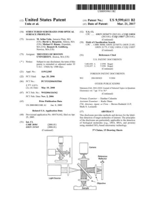
Structured substrates for optical surface profiling
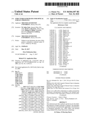
Structured substrates for optical surface profiling
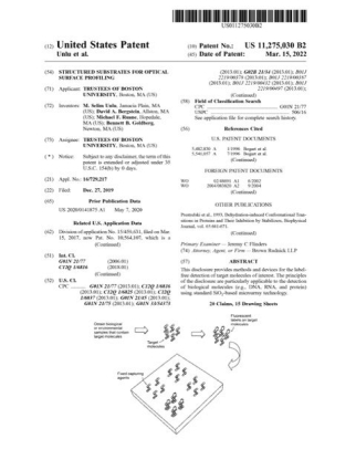
Structured substrates for optical surface profiling

Nanoparticles for self-referencing calibration
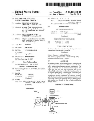
Polarization-enhanced interferometric imaging
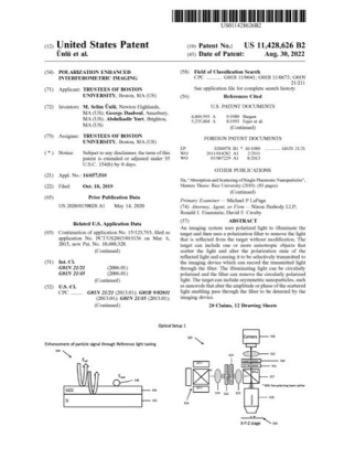
Polarization-enhanced interferometric imaging
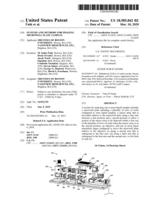
Imaging samples in microwell plates
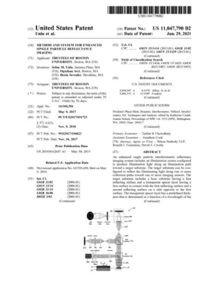
Enhanced single-particle reflectance imaging
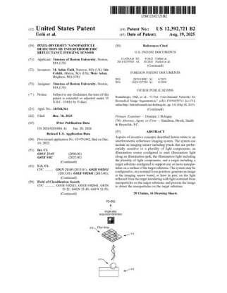
Pixel-diversity nanoparticle detection with the IRIS sensor
Dynamic tracking of captured targets for digital biosensing
Dynamic tracking of captured targets for digital biosensing
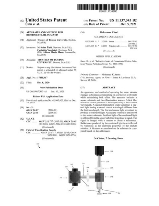
Apparatus and method for biomolecular analysis
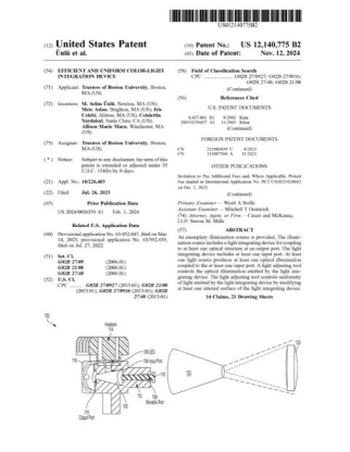
Uniform color-light integration device, the Euclid light source
Uniform color-light integration device, the Euclid light source
Pixel-diversity nanoparticle detection with the IRIS sensor
Advanced Interferometric Reflectance Imaging Sensor (IRIS) and uses thereof
All 16 as a list
| Patent | Title | Area | Status |
|---|---|---|---|
| US 9,599,611 B2 | Structured substrates for optical surface profiling | The sensor chip and its substrate | Issued 2017 |
| US 10,564,107 B2 | Structured substrates for optical surface profiling | The sensor chip and its substrate | Issued 2020 |
| US 11,275,030 B2 | Structured substrates for optical surface profiling | The sensor chip and its substrate | Issued 2022 |
| US 10,151,680 B2 | Nanoparticles for self-referencing calibration | The sensor chip and its substrate | Issued 2018 |
| US 10,488,328 B2 | Polarization-enhanced interferometric imaging | Imaging methods | Issued 2019 |
| US 11,428,626 B2 | Polarization-enhanced interferometric imaging | Imaging methods | Issued 2022 |
| US 10,585,042 B2 | Imaging samples in microwell plates | Imaging methods | Issued 2020 |
| US 11,047,790 B2 | Enhanced single-particle reflectance imaging | Single-particle and nanoparticle detection | Issued 2021 |
| US 12,392,721 B2 | Pixel-diversity nanoparticle detection with the IRIS sensor | Single-particle and nanoparticle detection | Issued 2025 |
| US 11,561,221 B2 | Dynamic tracking of captured targets for digital biosensing | Single-particle and nanoparticle detection | Issued 2023 |
| US 12,474,338 B2 | Dynamic tracking of captured targets for digital biosensing | Single-particle and nanoparticle detection | Issued 2025 |
| US 11,137,343 B2 | Apparatus and method for biomolecular analysis | The instrument | Issued 2021 |
| US 12,140,775 B2 | Uniform color-light integration device, the Euclid light source | The Euclid light source | Issued 2024 |
| US 18/909,517 | Uniform color-light integration device, the Euclid light source | The Euclid light source | Pending. Filed October 2024, divisional |
| US 19/276,607 | Pixel-diversity nanoparticle detection with the IRIS sensor | Single-particle and nanoparticle detection | Pending. Filed July 2025, continuation |
| PCT/US2026/037270 | Advanced Interferometric Reflectance Imaging Sensor (IRIS) and uses thereof | The instrument | Pending. Filed July 2026, international application |
Application notes and technical literature.
The worked experiments behind the platform, plus the spec sheets. Every document is a direct PDF download.
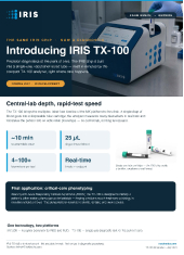
IRIS TX-100
TX-100 is the tabletop clinical instrument. In development, not available for sale.
Product literature, July 2026 IRIS_TX-100.pdf
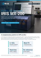
IRIS MX-200
MX-200 is the multiplex research instrument, successor to the MX-104. Spec sheet.
Product literature, July 2026 IRIS_MX-200.pdf
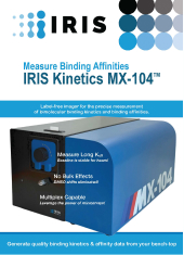
IRIS MX-104
MX-104 is the multiplex instrument shipping today. Sample volume, chamber, flow rates and cycle time.
Product literature, July 2025 MX-104-Jul2025.pdf

Closing the assay development gap
A Boston University and IRIS Kinetics review. Its own opening position is that IRIS is a bridge, not a replacement. Carries the IL-6 case study.
Technical review, 2026 Assay-Development-Gap.pdf
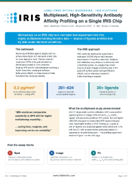
Multiplexed profiling in one run
More than thirty anti-LF camelid VHH ligands profiled in a single run, including one ligand that gave no measurable SPR signal.
Application note, July 2026 Mplex-July-2026.pdf
App Note IK-12: the diagnostic platform
Attomolar microRNA, allergen-specific IgE in whole blood and serum, and a method comparison table.
Application note, July 2025 App-Note-12-Diagnostic-Platform.pdf
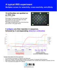
A worked multiplex panel
Ten antibodies spotted, four antigens injected in sequence, with detection antibodies. Two anti-IL-8 antibodies binding at different affinities.
Application note, July 2026 IL6-panel.pdf
Want to run your own?
The instruments behind this library are the ones we sell.
IRIS instruments are already in labs across the country empowering faster, better research.