Technology

IRIS stands for interferometric reflectance imaging sensor. It measures label-free biomolecular binding as a change in optical thickness on a silicon chip, read by the interference of reflected light.
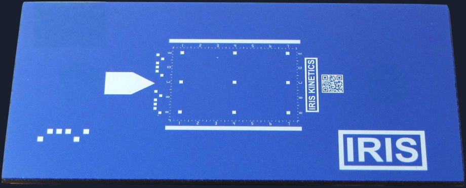
The IRIS chip, photographed from above: a blue 8 by 16 mm silicon chip with an alignment arrow, a scale grid, a QR code and the IRIS mark
The IRIS Chip: silicon under a single oxide layer, ready to be spotted with a capture microarray.

Precision interference.

Light reflects from the top of the chip’s oxide layer and again from the silicon beneath, and the two reflections interfere. Bound biomass thickens the film and shifts the reflectance curve. IRIS reads that shift independently of the solution’s refractive index, to better than pg/mm². SPR measures a change in refractive index and converts it to mass; IRIS measures the mass accumulating on the surface directly, which is why pg/mm² is routine for IRIS and hard for SPR.
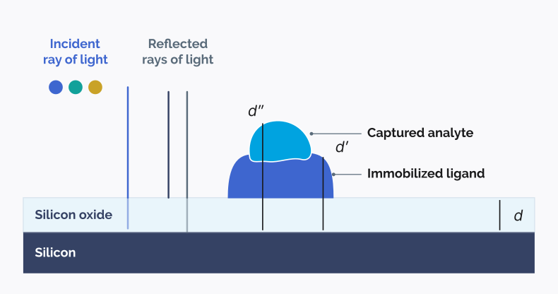
Cross-section of an IRIS chip. Incident light at three wavelengths falls on a silicon oxide layer over silicon and two rays reflect back. An immobilized ligand sits on the oxide and a captured analyte sits on the ligand. Three thickness marks show d for the bare oxide, d prime for oxide plus ligand, and d double prime for oxide plus ligand plus analyte.
An immobilized ligand raises the film’s optical thickness from d to d′; a captured analyte raises it again to d″. That difference is the readout.
White light dispersed by a prism.

From white light
to a readout.

Each wavelength reflects differently from a film of a given thickness. The instrument illuminates the chip at a set of chosen wavelengths and reads which of them return strongly. A thickening film shifts that pattern.

Versatile chip design.

Silicon oxide coated chips carry an immobilization layer and are spotted with capture molecules, most commonly antibodies, to produce multiplex capture microarrays. Tens to hundreds of probes per chip capture antigens, viruses and bacteria. Each chip is assembled into a flow cell and used once.

8 × 16 mm chip
Hundreds of spots per array
Single use
IRIS Chip specifications
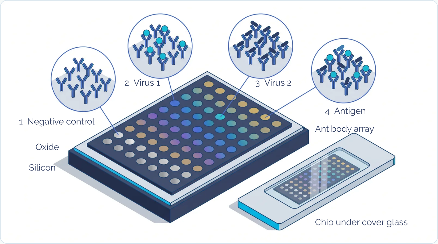
Isometric illustration of an IRIS chip: a grid of coloured antibody spots on a dark array surface, sitting on a pale oxide layer over a navy silicon base. Four call-outs show the spot types: a negative control antibody with nothing bound, an antibody with virus 1 particles bound, an antibody with virus 2 particles bound, and an antigen-specific antibody. A second chip sits beside it under cover glass.
Negative control antibody
Virus 1-specific antibody
Virus 2-specific antibody
Antigen-specific antibody
Each spot is one printed capture antibody: the control binds nothing, the virus-specific antibodies capture whole virus, the antigen-specific antibody captures viral antigen.

Real-time imaging.

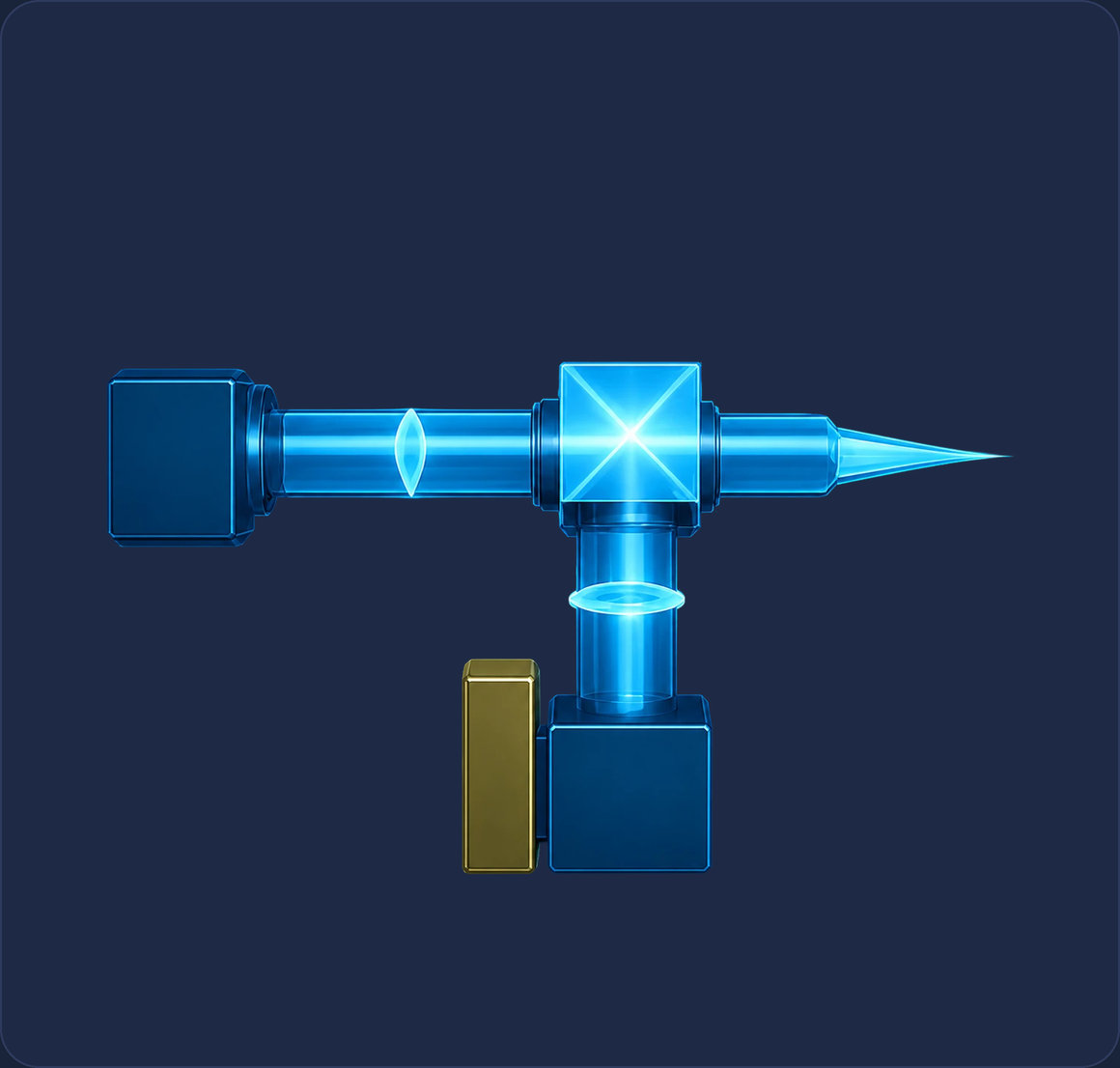
As sample flows over the chip inside the flow cell, the imaging system acquires full images of the array throughout the run. Four-color LEDs feed the Euclid™ light source, which flattens illumination across the chip; a CMOS camera records every spot in the same frame.
Live sensor output: each spot brightens as bound material accumulates.
The IRIS imaging system. Four-color LEDs feed the Euclid light source; the light passes a collimating lens and critical illumination into a beam splitter and a 2x objective, and the returning image passes a tube lens into a CMOS camera.
The imaging system, schematic. Our proprietary imaging system allows for much higher sensitivity measurements.

Continuous imaging yields the full kinetic trace.

Every spot is imaged throughout the run, giving a continuous record of bound mass against time.
The association rate (kon) and dissociation rate (koff) are fitted from that trace, and the affinity (KD) follows from their ratio.

<1

pg/mm² demonstrated surface mass sensitivity, rivalling SPR

Mplex, July 2026
6

pg/mm² is what 1 ng/mL of IL-6 puts on the surface, label-free

Assay Development Gap, 2026
10−6s−1

off-rates (koff) measured directly, not extrapolated

MX-104, July 2025

Backed by 18 years of published results.

IRIS instruments run in more than ten research institutions, and independent groups publish with them. The technology is also the subject of 25 PhD dissertations.

18

years continuously in print, 2008 to 2025

the publication record
3,472

citations across the published record

Google Scholar, checkable to the row
13

issued patents, from the light source to single-particle detection

the patent list